Research Stories
Bone Tissue Engineering via Application of a Collagen/Hydroxyapatite 4D-printed Biomimetic Scaffold for Spinal Fusion
Utilization of bioimplant medical device in a Bio-medical field
Bio-Mechatronic Engineering
Prof.
KIM, GEUNHYUNG
Hwangbo Hanjun, Dr. Hyeongin Lee
In an aging population, the frequency of patients with spine-related problems such as spinal stenosis, vertebral fractures, progressive deformities, and instability has shown a concerning increase. Due to the superior biological properties, biomimetic bone grafts have been long considered to be the reference standard for successful bone tissue regeneration and spinal fusion.
Prof. Geun Hyung Kim and his research team (Hanjun Hwangbo and Dr. Hyeongjin Lee; first author) from the department of biomechatronic engineering / biomedical institute for Convergence at SKKU (BICS) conducted joint research with Prof. In-Bo Han and his research team (Eun Ji Roh; first author) from CHA University School of medicine to develop 4D printing strategy to fabricate biomimetic microchanneled collagen/hydroxyapatite scaffold (MC- scaffold). The fabricated MC- scaffold was further analyzed in a spinal fusion model of a mouse. Consequently, the results indicate significantly higher bone regeneration and rate of spine fusion compared to the conventionally fabricated collagen/hydroxyapatite scaffold.
A major drawback of the conventionally used bone grafts is the limited vascular network causing insufficient nutrient and metabolite transfer to the surrounding tissue. To overcome this issue, Prof Geun Hyung Kim and his research team utilized a 4D printing strategy (one-way-shape-morphing) to fabricate a biomimetic microchanneled scaffold composed of collagen and hydroxyapatite (the main component of native bone) to induce a higher degree of osteogenesis and angiogenesis. These findings are owed to the synergistic effects of high infiltration of blood vessels into the microchannels and superior biophysical properties of the microchanneled collagen/hydroxyapatite scaffold.
Additionally, Prof. Geun Hyung Kim and his research team (WonJin Kim; first author) collaborated with Prof. SangJin Lee and his research team (Dr. Hyeongjin Lee; first author) from Wake Forest Institute for Regenerative Medicine (WFIRM) applied 4D printing strategy to induce cellular alignment in extracellular matrix (ECM) based scaffold. Subsequently, a significantly higher degree of muscle regeneration was observed via implantation of the 4D printed bioconstruct into a muscle defect in the rat model.
Currently, mimicking the uniaxial alignment of muscle cells as presented in the native skeletal muscle is often a challenging and difficult task to accomplish with conventional 3D printing. To overcome this issue, Prof. Geun Hyung Kim and his research team utilized developed a composite bioink composed of natural polymer (ECM) and synthetic polymer (Polyvinyl alcohol, PVA), and a 4D printing strategy to provide topographical cue in ECM-based scaffold to induce cellular alignment of primary human muscle progenitor cell (hMPC). Various process parameters such as PVA molecular weight and pneumatic pressure have been analyzed to optimized to obtain the best biological factors such as cellular alignment and differentiation of the host cells.
In addition, to conduct in vivo investigations into muscle regeneration, the bioconstruct sized 15 × 7 × 3 mm3 have been printed and implanted into the tibialis anterior (TA) muscle defect. The cell viability of the fabricated bioconstruct was measured to be around 90%. After 8 weeks of implantation, the muscle regeneration on rats that received the 4D bioprinted structure was significantly higher compared to the conventional 3D printed structure.
The presented 4D printing strategy enables versatility of designing various biological and physical factors of the implant grafts for effective tissue regeneration. Moreover, the introduced 4D printing strategy is believed to have multiple applications such as tendon, nerve, or cardiac.
These studies were supported by a grant from the National Research Foundation of Korea (NRF) funded by the Ministry of Education, Science, and Technology and by an NRF grant funded by the Ministry of Science and ICT for the Bioinspired Innovation Technology Development Project. In addition, both studies were published in “Applied Physics Reviews” (IF = 17.05) and were selected as a featured paper.
Figure 1. Schematical illustration and SEM images of microchanneled collagen/HA scaffold.
Figure 2. Immunofluorescent images of the retrieved TA muscles for MRPL12 (green)/MHC (red) at 8 weeks after implantation.
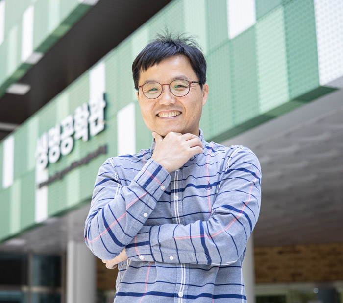
Professor Geun Hyung Kim
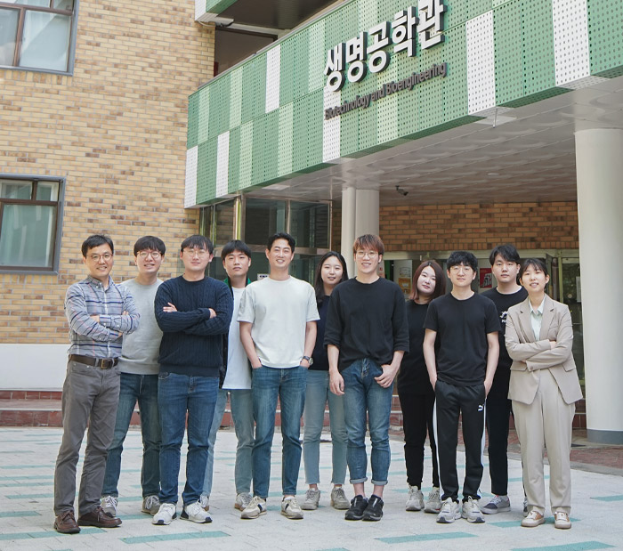
Professor Geun Hyung Kim and his research team
(From right) Prof. Geun Hyung Kim, Dr Hyeongjin Lee, Mr WonJin Kim, Mr Hanjun Hwangbo


